4 Stages of Appendicitis
The Four Stages of Appendicitis: A Comprehensive Medical Guide
Appendicitis represents a common yet potentially serious medical condition that requires prompt recognition and treatment. This comprehensive guide provides detailed information about the four distinct clinical stages of appendicitis, offering clarity on symptom progression, diagnostic considerations, and appropriate medical responses. Understanding these stages in their complete context empowers individuals to make informed healthcare decisions and seek timely medical intervention when necessary.
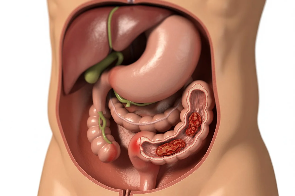
The appendix, a small tubular structure located in the lower right abdomen, can become inflamed due to various factors including obstruction, infection, or other pathological processes. This inflammation progresses through predictable stages, each with distinct clinical features and implications for treatment approaches. Early recognition of symptoms and appropriate medical evaluation can significantly influence treatment outcomes, recovery timelines, and potential complication rates associated with appendicitis.
Understanding Appendix Anatomy and Physiological Function
The appendix measures approximately 2-4 inches in length and is attached to the cecum, which represents the beginning portion of the large intestine. While its exact physiological function continues to be studied in medical research, current scientific understanding suggests it may play a role in immune system function and gut microbiome maintenance. Despite its potential physiological roles, the appendix remains susceptible to inflammation that can progress through the characteristic stages of appendicitis, requiring medical attention.
Appendiceal inflammation typically begins with obstruction of the appendiceal lumen. This obstruction may result from various factors including fecal material accumulation, lymphoid hyperplasia, foreign bodies, or rarely, tumors. The resulting closed-loop obstruction leads to bacterial overgrowth, increased intraluminal pressure, and compromised blood flow—initiating the inflammatory cascade that defines appendicitis progression through its subsequent stages that we will explore in detail.
The Four Clinical Stages of Appendicitis Development
Appendicitis progression follows a relatively predictable timeline through four distinct clinical stages. Each stage represents increasing severity of inflammation and tissue compromise, with corresponding implications for diagnosis, treatment urgency, and potential complications. Medical professionals use this staging system to guide diagnostic and therapeutic decision-making, ensuring appropriate management based on disease severity and progression.
The complete understanding of these four stages provides valuable insight into symptom patterns, expected timelines, and appropriate medical responses. This knowledge is particularly valuable for recognizing early warning signs, understanding when emergency care becomes necessary, and appreciating the potential consequences of delayed treatment in appendicitis cases that progress through advanced stages.
| Stage | Clinical Name & Timeline | Clinical Features & Pathophysiology | Medical Management & Urgency |
|---|---|---|---|
| 1 | Early Appendicitis (0-24 hours from symptom onset) |
Periumbilical or epigastric discomfort progressing to anorexia and nausea. Appendiceal mucosa inflammation with luminal distension occurs. Minimal systemic symptoms are typically present during this initial phase of appendicitis development and progression. | Prompt clinical evaluation recommended with medical consultation. Diagnostic imaging may be considered based on clinical presentation. Antibiotic therapy may be initiated, with surgical consultation for potential appendectomy depending on assessment findings and patient factors. |
| 2 | Suppurative Appendicitis (24-48 hours from symptom onset) |
Pain localizes to right lower quadrant at McBurney’s point specifically. Fever, leukocytosis, and peritoneal signs develop clinically. Transmural inflammation with purulent exudate formation characterizes this stage of progressive appendiceal inflammation and infection development. | Emergency department evaluation required without delay. Surgical intervention typically indicated based on clinical and diagnostic findings. Intravenous antibiotics and fluid resuscitation initiated preoperatively to optimize patient condition before surgical management procedures. |
| 3 | Gangrenous Appendicitis (48-72+ hours from symptom onset) |
Intense localized pain with possible temporary relief due to nerve ending compromise. Systemic toxicity with fever, tachycardia, and clinical deterioration. Tissue ischemia and necrosis develop with compromised vascular supply to appendiceal structures at this advanced stage. | Immediate surgical consultation mandatory without exception. Broad-spectrum antibiotics essential for infection control. Risk of perforation elevated significantly, requiring urgent operative management to prevent catastrophic complications and systemic infection spread. |
| 4 | Perforated Appendicitis (72+ hours from symptom onset typically) |
Generalized peritonitis with rigid abdomen and diffuse tenderness. High fever, significant leukocytosis, systemic inflammatory response syndrome. Appendiceal rupture with intra-abdominal contamination and potential abscess formation characterizes this final emergency stage. | Surgical emergency requiring immediate intervention without delay. Extended antibiotic therapy and possible peritoneal drainage procedures necessary. Increased complication rates and longer hospitalization expected with more complex postoperative recovery management required. |
Stage 1: Early Appendicitis (0-24 Hours from Symptom Onset)
The initial stage of appendicitis typically presents with visceral, poorly localized pain that often begins in the periumbilical or epigastric regions. This pain results from distension of the appendiceal lumen and stimulation of visceral nerve fibers that respond to stretching and pressure changes within the intestinal tract. Accompanying symptoms frequently include anorexia (loss of appetite) and mild nausea, though vomiting is less common in this early phase of appendicitis development.
Pathologically, early appendicitis involves mucosal inflammation and edema with luminal distension characteristic of the initial inflammatory response. The appendix remains intact without transmural inflammation or significant compromise to its blood supply during this stage. Physical examination may reveal mild tenderness without definitive localization, and systemic signs such as fever are typically absent or minimal during this initial stage of appendicitis development, making clinical diagnosis challenging at times.
Stage 2: Suppurative Appendicitis (24-48 Hours from Symptom Onset)
As inflammation progresses through the stages of appendicitis, pain characteristically migrates to the right lower quadrant, localizing specifically at McBurney’s point—approximately one-third the distance from the anterior superior iliac spine to the umbilicus anatomically. This shift represents progression from visceral to parietal pain as inflammation extends to involve the parietal peritoneum, which has more precise localization capabilities in pain perception.
The appendix becomes markedly inflamed with purulent exudate formation, and transmural inflammation develops throughout the appendiceal wall. Systemic signs emerge clinically, including fever (typically 37.7-38.3°C), leukocytosis with elevated white blood cell counts, and tachycardia. Physical examination demonstrates localized tenderness with possible rebound tenderness and guarding indicative of peritoneal irritation. Diagnostic accuracy increases during this stage with characteristic findings on physical examination and imaging studies that help confirm appendicitis diagnosis.
Stage 3: Gangrenous Appendicitis (48-72+ Hours from Symptom Onset)
Gangrenous appendicitis represents progression to tissue necrosis resulting from compromised vascular supply to the appendix. The inflammatory process causes thrombosis of appendiceal vessels, leading to ischemic necrosis of the appendiceal wall with potential bacterial translocation. A potentially misleading clinical sign is temporary pain relief as necrotic tissue loses neural function and sensation capability, which may be misinterpreted as improvement by patients.
Systemic toxicity becomes more pronounced with higher fever (often exceeding 38.3°C), significant leukocytosis with left shift, and possible tachycardia indicating systemic inflammatory response. The patient appears clinically ill, and abdominal examination reveals marked localized tenderness with definite peritoneal signs including rigidity and rebound tenderness. The risk of perforation increases substantially during this stage, necessitating urgent surgical intervention to prevent catastrophic complications associated with appendiceal rupture.
Stage 4: Perforated Appendicitis (72+ Hours from Symptom Onset)
Perforation represents the most advanced stage of appendicitis, occurring when necrotic appendiceal wall integrity fails completely, releasing purulent and fecal material into the peritoneal cavity. This contamination causes diffuse peritonitis with generalized abdominal pain, rigidity, guarding, and systemic inflammatory response that can progress to sepsis without prompt intervention.
Clinical presentation includes high fever, significant leukocytosis often exceeding 15,000 cells/mm³, tachycardia, and possible hypotension in severe cases indicating developing septic shock. Abdominal examination demonstrates generalized tenderness, rebound tenderness, and involuntary guarding throughout the abdomen. Complications increase substantially at this stage, including abscess formation, sepsis syndrome, prolonged recovery, and increased morbidity. Immediate surgical intervention with broad-spectrum antibiotic coverage is mandatory for survival and optimal outcomes.
Clinical Pearl: The migration of pain from periumbilical to right lower quadrant represents a classic diagnostic feature of appendicitis that aids in clinical recognition. However, approximately one-third of patients may not exhibit this classic pattern due to anatomical variations. Anatomical variations, including retrocecal or pelvic appendix positions, can alter pain presentation and localization, requiring heightened clinical suspicion and appropriate diagnostic imaging for accurate diagnosis.
Diagnostic Considerations and Comprehensive Medical Evaluation
Accurate diagnosis of appendicitis requires integration of clinical history, physical examination findings, and appropriate diagnostic studies in a systematic approach. The Alvarado scoring system (MANTRELS criteria) provides a validated clinical prediction tool incorporating migration of pain, anorexia, nausea/vomiting, tenderness in right lower quadrant, rebound pain, elevated temperature, leukocytosis, and shift to left in white blood cell differential. A score ≥7 suggests high probability of appendicitis requiring further evaluation.
Laboratory evaluation typically includes complete blood count with differential (demonstrating leukocytosis with neutrophil predominance) and C-reactive protein measurement as inflammatory markers. Urinalysis helps exclude urinary tract pathology that might mimic appendicitis symptoms. Imaging modalities include ultrasound (particularly valuable in children and pregnant patients due to absence of radiation) and computed tomography with sensitivity and specificity exceeding 90% in most clinical studies. Diagnostic laparoscopy may be considered in uncertain cases when imaging is inconclusive.
Therapeutic Management Approaches and Treatment Considerations
Surgical appendectomy remains the definitive treatment for acute appendicitis across most clinical presentations. Laparoscopic approach offers advantages including reduced postoperative pain, shorter hospitalization duration, faster recovery to normal activities, and improved cosmetic outcomes compared to open appendectomy techniques. However, open approach may be preferred in cases of perforation with diffuse peritonitis or when laparoscopic expertise is limited based on institutional resources.
Antibiotic therapy plays a crucial adjunctive role in appendicitis management. Preoperative antibiotics reduce surgical site infections significantly, while postoperative therapy duration depends on operative findings and severity of infection encountered. Non-operative management with antibiotics alone may be considered for selected patients with uncomplicated appendicitis and specific clinical factors, though approximately 20-30% experience recurrence within one year, necessitating eventual appendectomy in many cases.
Frequently Asked Questions About Appendicitis
Comprehensive Conclusion and Summary
Understanding the four stages of appendicitis provides essential knowledge for recognizing symptom progression and seeking appropriate medical care promptly. Early diagnosis during initial stages allows for straightforward surgical management with minimal complications and rapid recovery typically. Delayed recognition increases risks of perforation, peritonitis, and complex postoperative courses significantly, highlighting the importance of timely medical evaluation for abdominal pain.
Clinical vigilance remains paramount, particularly for high-risk populations including pediatric, elderly, and pregnant patients who may present with atypical symptoms requiring heightened suspicion. Integration of clinical assessment with appropriate diagnostic imaging optimizes diagnostic accuracy while minimizing unnecessary procedures. Surgical appendectomy, increasingly performed laparoscopically, continues as the definitive treatment for most cases, with antibiotic therapy playing an important adjunctive role in comprehensive management of appendicitis across its various stages.


